
Finland: A team of researchers at Tampere University has developed a new generation of 3D-printed bone implants that could transform the way damaged bones are repaired, offering a safer and more personalized alternative to traditional bone grafting procedures.
Bone grafting is currently the world’s second most common tissue transplantation procedure, with more than two million surgeries carried out every year.
Existing treatments often depend on bone taken either from the patient’s own body or from donors, methods that can lead to additional surgeries, longer recovery periods, and limited availability of suitable graft material.
The new research, led by Dr. Antonia Ressler, Postdoctoral Research Fellow at the Tampere Institute for Advanced Study, focuses on creating synthetic bone-like scaffolds using hydroxyapatite—the same mineral compound naturally found in human bones.
By combining hydroxyapatite with advanced ceramic 3D-printing technology, the researchers succeeded in producing implants that closely imitate the structure of real bone. The implants can also be individually designed to match a patient’s exact bone defect, improving precision in treatment.
Dr. Antonia Ressler, lead researcher of the project, said that, “Using the same material found in natural bone allows us to create implants that work with the body’s own healing process. At the same time, ceramic 3D printing enables us to tailor each implant according to the patient’s needs without relying on drugs or growth factors that may produce side effects.”
The breakthrough is the result of four years of work under the AffordBoneS project, supported by the Horizon Europe Marie Skłodowska-Curie Postdoctoral Fellowship program.
Researchers are now continuing the work through a follow-up initiative called GlassBoneS, which aims to make advanced bone regeneration treatments more affordable and widely accessible.
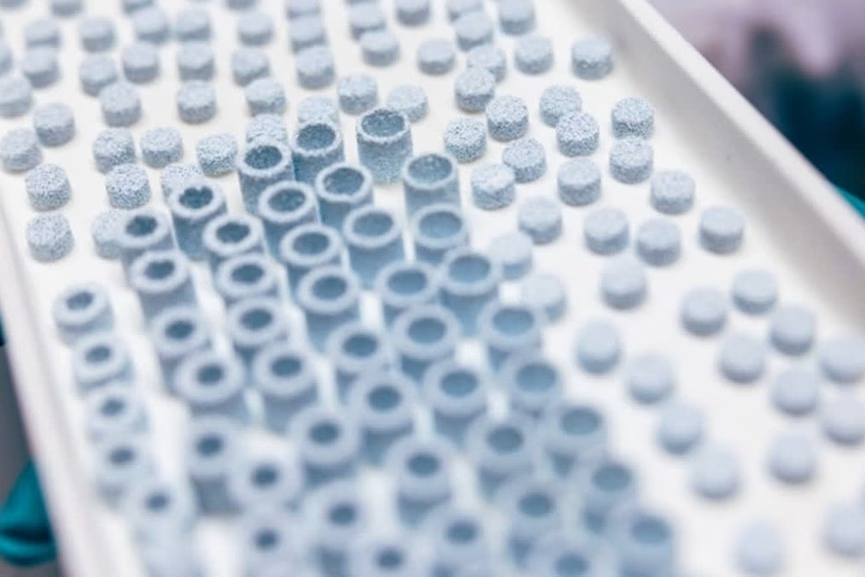
Using ceramic 3D printing, the team carefully engineered the internal structure of the implants, including the size and arrangement of microscopic pores that allow cells, oxygen, and nutrients to move through the scaffold.
The researchers identified an ideal design featuring pores measuring around 400 micrometers with nearly 45 percent porosity.
According to the team, this structure provided the right balance between mechanical strength and biological performance, enabling bone-forming cells to attach, communicate, and begin generating new tissue.
The study also revealed that the surface quality of the material plays a major role in successful healing. Researchers found that extremely high manufacturing temperatures can alter the surface of the implants, making it harder for human cells to attach properly.
The findings could mark an important step toward more reliable and patient-specific bone regeneration therapies, particularly as aging populations worldwide continue to increase demand for orthopedic and reconstructive treatments.